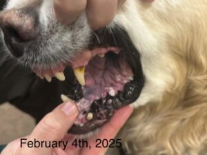
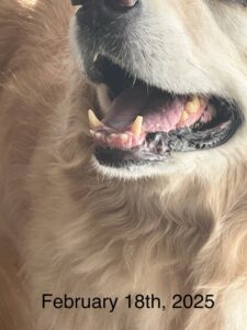
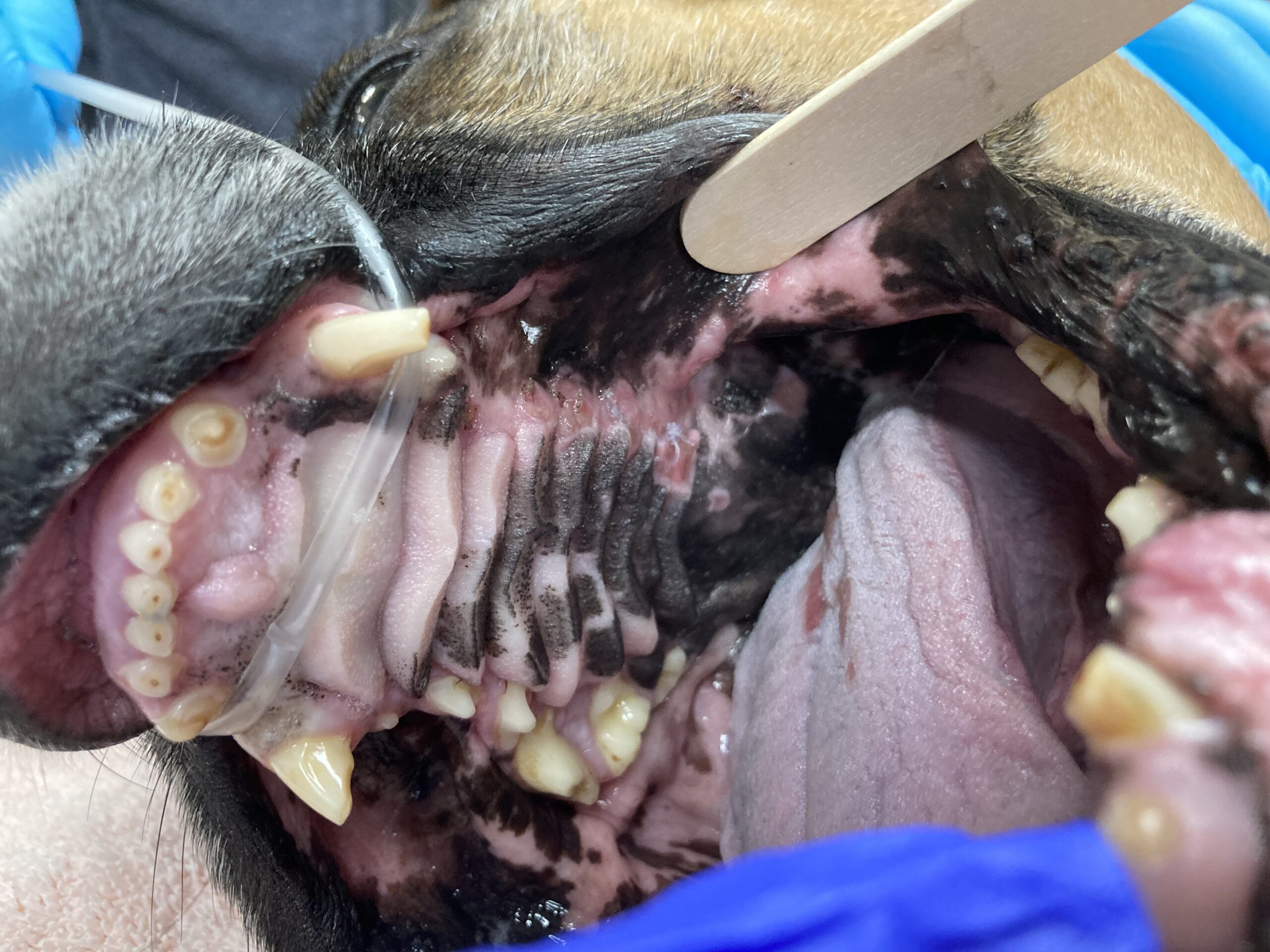
Granger was an 11 y FS DMH who presented to our Internal Medicine service for weight loss and nasal signs. Her owners reported that her sneezing, congestion, and loud breathing had gotten progressively worse over the past year. In the past few months she had a few episodes of bloody nasal discharge and sneezing fits. She became very picky about which foods she would eat at home. She had previously been placed on oral antibiotics for suspected chronic rhinitis, but they had only temporarily improved her clinical signs.

On physical examination she has a thin body condition, was stertorous, and had evidence of nasal congestion. A CBC, chemistry, and T4 were unremarkable. Thoracic radiographs revealed only aerophagia (suspected to be secondary to severe nasal disease) but was otherwise unremarkable. An abdominal ultrasound was unremarkable. Granger was admitted to the hospital for a head CT and rhinoscopy with Dr. Sarah Noble for continued investigation into the cause of his clinical signs.

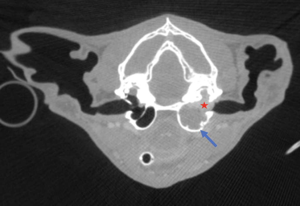
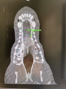
Her head CT revealed a large peripherally contrast-enhancing and irregularly marginated ovoid mass filling the nasopharynx and middle ear. There were multiple areas of lysis associated with the osseous bulla, and secondary obstructive rhinitis was suspected. The medial retropharyngeal lymph nodes were mildly enlarged, which was suspected to be due to reactive hyperplasia. Differentials for this type of mass on CT include carcinoma vs fungal disease (cryptococcus) vs a chronically infected nasopharyngeal polyp with concurrent otitis media/interna.

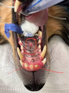
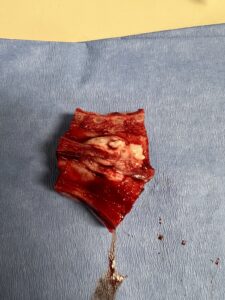
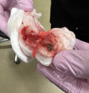
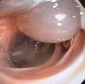
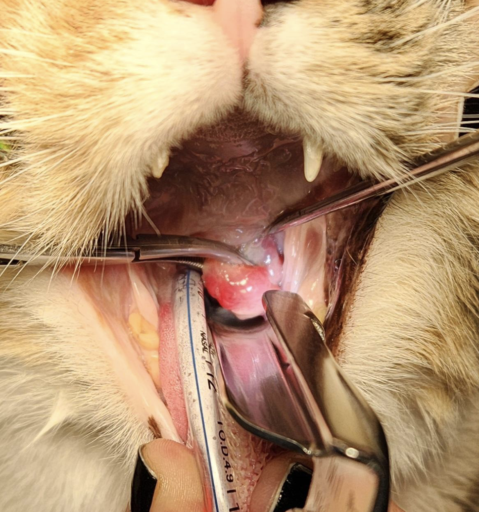
Dr. Noble removed the large, irregular looking mass from the nasopharynx under general anesthesia. A polyp vs neoplasia was prioritized based on the appearance of the mass. There was mild bleeding with some draining purulent material following mass removal, but Granger’s recovery from general anesthesia was uneventful. The mass was submitted for histopathology, and the pathologist confirmed her mass was an “ulcerated and severely inflamed benign feline nasopharyngeal polyp”. As of the publication of this article she has continued to do well at home.

CLINICALLY RELEVANT FACTS:

  • Feline nasopharyngeal polyps should be considered a differential for chronic nasal signs in cats, despite their age.
  • In cat’s with this condition surgical removal can be curative, however regrowth of the mass occurs in some patients which may require additional treatment. A ventral bulla osteotomy will often be recommended in these cases. It has previously been reported in the literature that 50% of polyps will regrow in feline patients.
  • Removal of the polyp will often lead to Horner syndrome (oculosympathetic dysfunction) on the side of the polyp’s origin. Owners should be warned about the potential of polyp regrowth, as well as the risk of Horner syndrome which may not resolve.

The red star shows the contrast enhancing mass seen in the left nasopharynx and middle ear. The left osseous bulla (blue arrow) is thin, expanded compared to the right, and has areas of bony lysis.

The nasopharyngeal polyp is visible as Dr. Noble applies gentle traction.

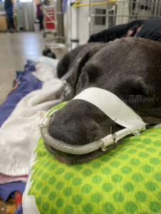
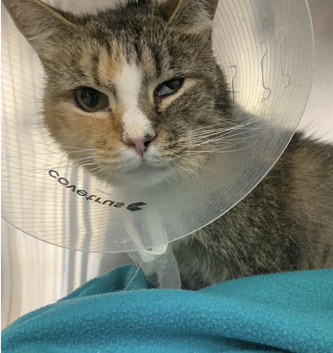
Granger had left Horner syndrome (left ptosis, miosis, enophthalmos, and third eyelid elevation) following the procedure.

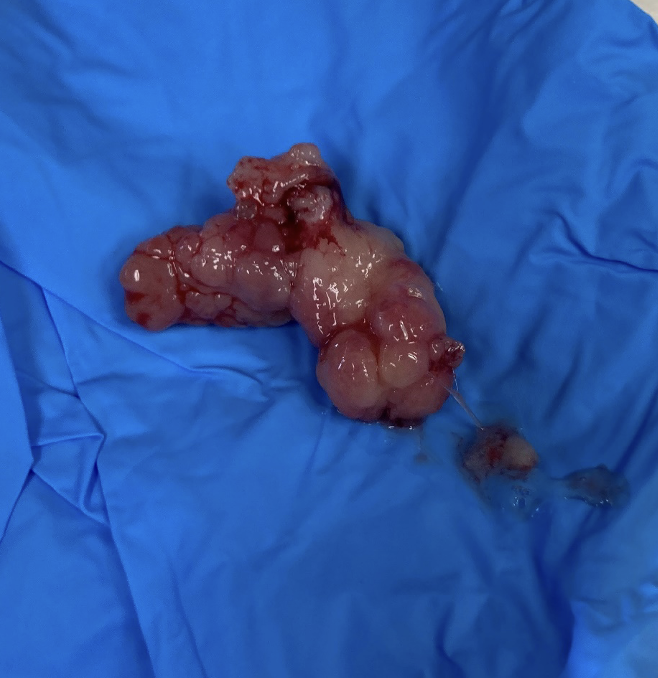
The irregularity of the mass was concerning for neoplasia, but was ultimately due to chronic ulceration and inflammation.

Authored by: Dr. Landry, DVM, DACVIM, Neurology 


Lucy was presented to our Oncology service for consultation following a diagnosis of large cell lymphoma by her primary care veterinarian. In June 2023, Lucy was presented to her primary care veterinarian for lethargy and left-sided facial swelling. The owner elected to treat empirically for an infectious/inflammatory process with antibiotics and a non-steroidal anti-inflammatory medication. Lucy initially had a favorable response to empirical therapy, but the swelling returned in October 2023. Lucy was evaluated by her primary care veterinarian who appreciated generalized peripheral lymphadenopathy during her exam. Bloodwork revealed a non-regenerative anemia (26.1%), lymphocytosis (8.97K), monocytosis (3.18K), thrombocytopenia (97K) and elevated ALP (459). Cytology of her lymph node was most consistent with a diagnosis of large cell lymphoma and she was referred to PVESC at that time.

During her initial oncology consultation at PVESC, Lucy was treated with L-asparaginase to help control her disease while the owner considered the treatment options discussed. Unfortunately, due to various circumstances, flow cytometry could not be submitted for immunophenotyping. Lucy had a favorable response to L-asparaginase and the owner elected to move forward with single-agent doxorubicin therapy and Lucy received her first dose on 10/31/23. On 11/27/23, following two doses of doxorubicin, the owner reported enlarged lymph nodes. 

Cancer relapse was confirmed when Lucy returned to PVESC for her recheck on 12/7/23. The owner elected to continue treatment with the LOPP protocol. Lucy developed a grade 4 non-febrile neutropenia following her first treatment with the LOPP protocol and her lymph nodes continued to progress. Tanovea was recommended, but was cost prohibitive to the client. The client was strongly considering transitioning Lucy to hospice-care at her recheck on 12/28/23. Laverdia was discussed as a more affordable and potentially effective treatment option for Lucy and the owner consented to further treatment with it. 

Laverdia (Laverdia-CA1) is the first conditionally approved oral tablet to treat lymphoma in dogs. It works by selectively targeting and binding to transport proteins to block them from transporting tumor suppressor proteins out of the nucleus where they can no longer be useful. The end result is an abundance of tumor suppressor proteins within the cell nucleus and ultimately cell death. Treatment is administered at-home twice weekly and the dose should be escalated after two weeks if well-tolerated. The most common side effects include anorexia, vomiting, diarrhea, weight loss and lethargy. 

Treatment with Laverdia was initiated on 12/28/24. Lucy was rechecked monthly after starting treatment. She experienced grade 1-2 inappetence when her dose was escalated after two weeks, which responded favorably to treatment with ondansetron given 30-60 minutes before Laverdia. Lucy’s lymph nodes continued to decrease in size and were nearly normal in size by early March 2024. She continued to have a strong partial response until early August 2024 when progressive lymphadenopathy was noted during her recheck exam. 

Patients that respond poorly to initial chemotherapy often fail to achieve durable remission times when treated with rescue chemotherapy protocols. In Lucy’s case, despite a poor response to initial therapy, her response to Laverdia was strong (near remission) and durable (8 months)! Altogether, treatment bought Lucy an additional 10 beautiful months to spend with her family and live life to the fullest. 

Clinically Relevant Facts:

  • There are many different options for treating canine large cell lymphoma beyond standard of care CHOP therapy, including single-agent chemotherapy drug protocols and Laverdia.
  • Laverdia (verdinexor) is an FDA conditionally approved oral targeted drug for the treatment of canine lymphoma. Unlike prednisone, it does not appear to induce chemotherapy resistance and may be considered for patients awaiting their oncology consultation. 
  • Despite large T-cell lymphoma carrying a worse overall prognosis, the overall response rate to Laverdia is higher in dogs with T-cell lymphoma (71%) compared to dogs with B-cell lymphoma (38%), even in a relapse setting. 
  • Thus, Laverdia should also be considered for patients who have failed initial standard of care therapy and may yield unexpected outcomes.

Authored by: Brittanie Partridge, DVM, PhD, DACVIM (Oncology)


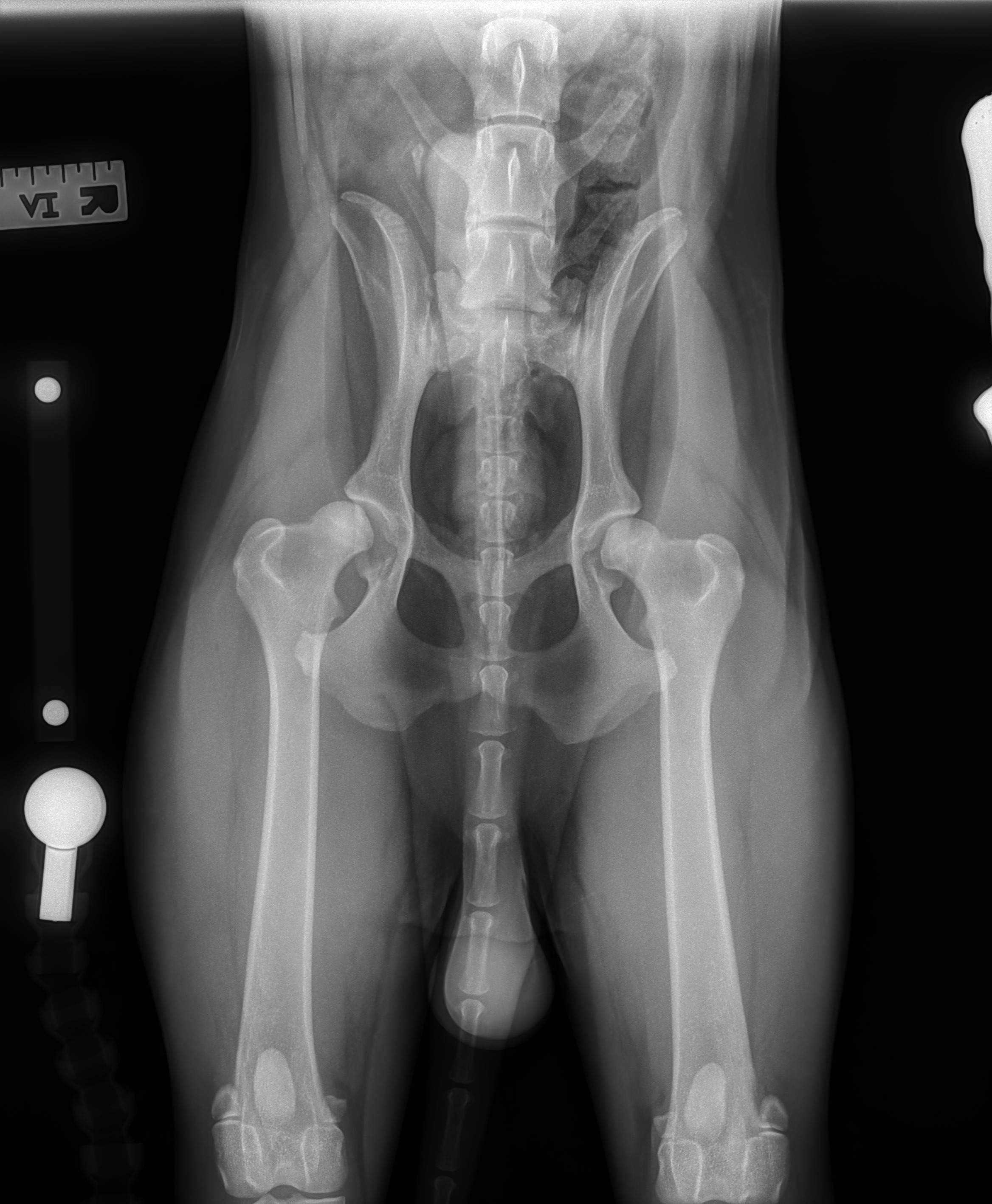
Arlo is 2.5-year-old male neutered Barbet from Canada who presented to Dr. Meghan Sullivan of the PVESC Surgery Department. He was diagnosed at 10 months of age with severe hip dysplasia. His owners noted that he would not jump onto anything and that his hips clicked when he walked. Arlo made the trip to PVESC at 12 months old and had templating radiographs (four views) performed to measure him for specific implant sizes. On examination, Arlo was walking well but had a stiffened hind end gait. Both hips were painful and had decreased range of motion bilaterally. There was also a loud clunking noise on range of motion of the right hip with significant subluxation.

Figure 1. VD pelvis showing significant hip dysplasia bilaterally with secondary degenerative changes prior to a year of age

Under sedation, he had significant Ortolani bilaterally. Radiographs revealed severe subluxation of both hips secondary to hip dysplasia. There was already mild degenerative joint changes as well as flattening of the femoral head, shallow acetabulum and thickening of the femoral neck bilaterally.

Arlo had his left total hip replacement performed at 13 months of age. Surgery went very well and he was weight bearing with sling assistance on the surgical leg later that evening. He had a BFX (biologic fixation system) which is a press fit (non-cemented) total hip replacement. He stayed overnight for 2 nights and was discharged with strict instructions for postop care. Recovery entailed 3 months of no off-leash and no access to furniture. He was allowed to go for short leash walks several times daily with sling assistance or help ‘em up harness. Arlo did great and his owners were perfectly compliant. Arlo had several weeks of NSAID’s, antibiotics, pain medications and several months of sedatives to aide in his activity restrictions.

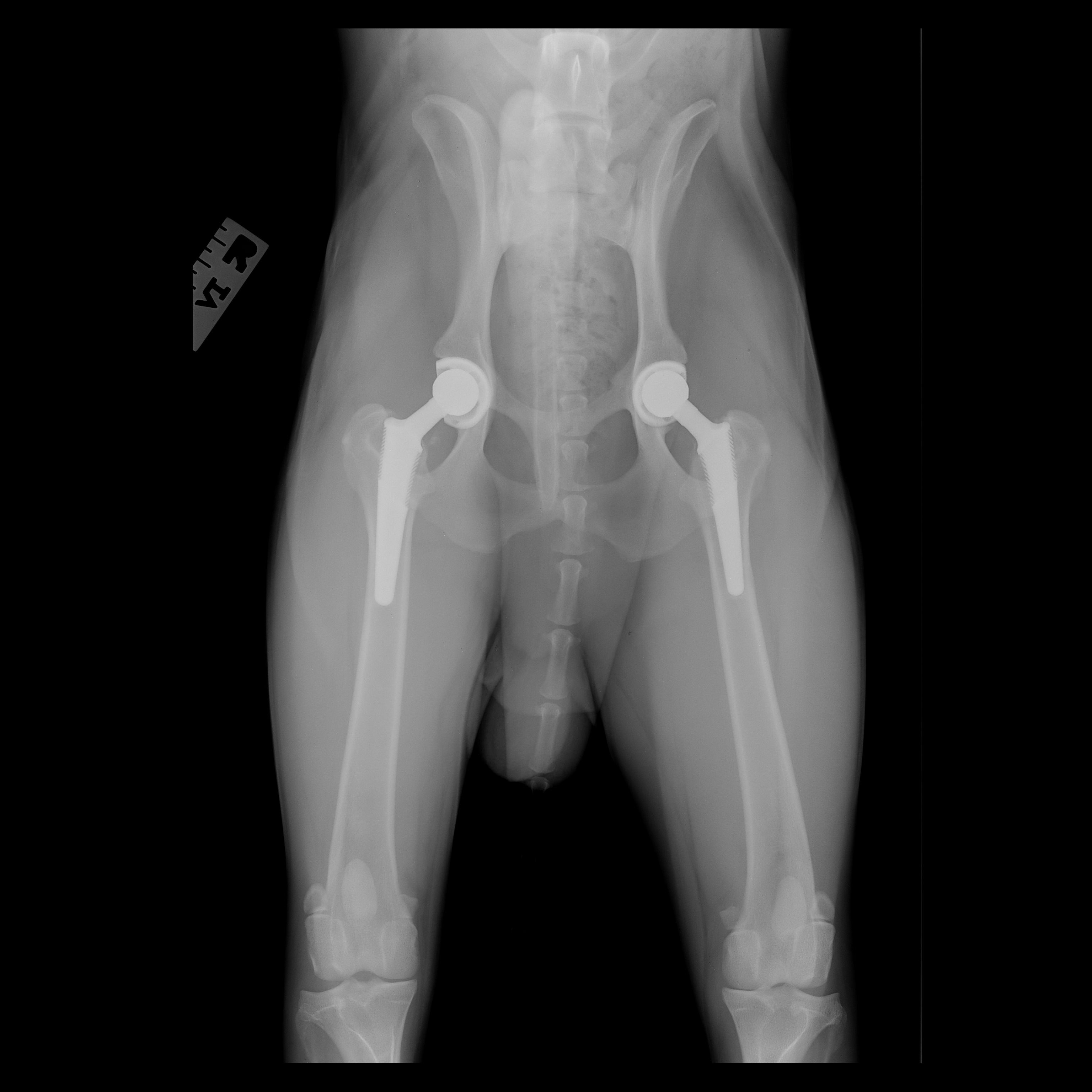
Arlo had a routine two-month recheck visit, and the radiographs showed a successful left total hip replacement. He was scheduled for his right total hip replacement one month later (three months after his left total hip replacement). The right total hip was successful with excellent anesthesia and an uneventful recovery. He was discharged similarly after two nights of 24/7 care and headed home to Canada to recover. He had another beautiful recovery, and recheck radiographs at two months showed routine healing. He is now a year out from his surgeries and running, playing, doing zoomies, hiking, etc. Arlo is having a blast now, being super active, happy, and living a pain-free life.

Figure 2. Recheck radiographs 2 months post final surgery: showing successful stages bilateral total hip replacement with BFX system

Total hip replacement is an option that delivers better functionality than the FHO (femoral head osteotomy). This procedure can be performed as early as 8-9 months of age, and consultation is best as soon as hip dysplasia is recognized to help achieve the best outcome. Dr. Sullivan at PVESC is the only surgeon in Maine performing total hip replacement surgery for dogs. We look forward to helping many more pets. To hear more about how to manage hip dysplasia and learn about all surgical options available to help them live their best lives, contact us today.

Authored by: Meghan Sullivan, DVM, DACVS-SA (Surgery)


Patient was a 13yr collie mix who has already undergone a splenectomy for a benign mass in 2022 and an adrenalectomy for an adenoma in 2023.

She presented in the summer of 2024 for neck pain and difficulty walking.  After testing positive for tick borne bacterial diseases Lyme, Anaplasma, and Rocky Mountain Spotted Fever, she was started on Doxycycline.

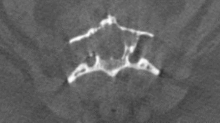
When her status did not improve, she was evaluated with a CT scan and found to have an extradural mass dorsal at C3 with bone lysis and spinal cord compression. Prednisone and Gabapentin were started.

Figure 1: CT scan of the patient 

As she continued to have neck pain and trouble walking, the owners elected to pursue surgery. A dorsal laminectomy was performed by Dr. Agrodnia and all visible compressive tissue was removed from above C3. Patient recovered well and was walking better within the week. She was referred to medical oncology who confirmed the diagnosis of plasma cell tumor and started appropriate medical management.

Authored by: Marta Agrodnia, DVM, DACVS-SA (Surgery)


Milo is an 11-year old MN Pomeranian who presented to our Neurology service for an acute onset of seizure-like episodes.

During the first episode, he began shaking, staring off, and holding up his right forelimb. He was then ataxic in all four limbs, quieter than normal for several minutes, and developed increased attention-seeking behavior with his owner for a few days.

On presentation to our Neurology service, Milo had an unremarkable neurological examination. Infectious testing revealed he was low positive for Rocky Mountain Spotted Fever (@ 1:25) and Anaplasma (@1:100), and he was started on a 4-week course of doxycycline. When Milo had another episode despite being on antibiotics (this time with a component of hypermetria prior to holding up his right forelimb), a brain MRI was scheduled to further investigate the cause of his signs.

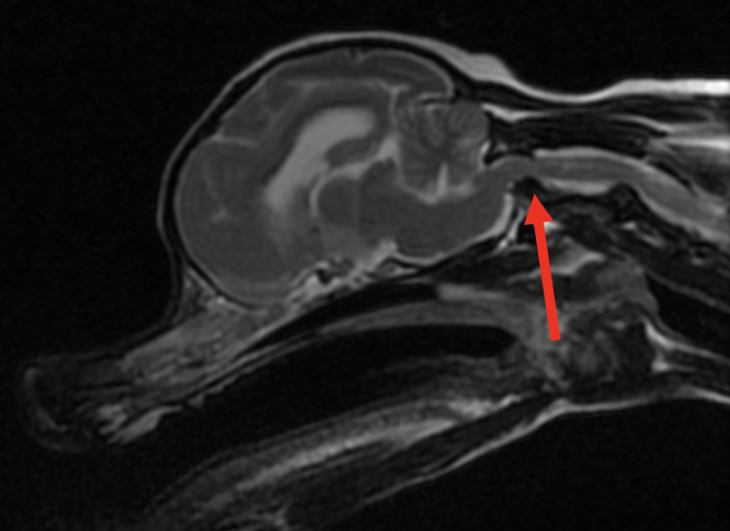
A brain MRI revealed a structurally unremarkable brain and atlantoaxial subluxation causing moderate extradural spinal cord compression.  This unexpected finding of vertebral instability is suspected to be the cause of Milo’s episodes. A cervical splint was placed to stabilize the site prior to discharge, which will require weekly changing for 8 weeks.

Figure 1. Sagittal T2- weighted image of the brain and high cervical spinal cord

The red arrow illustrates where the cranial aspect of C2 is dorsally displaced into the vertebral canal causing moderate extradural spinal cord compression and displacement. There is a loss of CSF and epidural fat signal at this site, consistent with spinal cord compression.

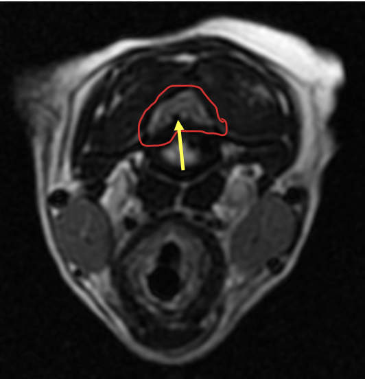
Figure 2. Axial T2- weighted image at the level of the cranial cervical spinal cord (red arrow in Figure 1)

The solid red line outlines the spinal cord which is moderately compressed from a protruding hypoplastic dens (yellow arrow).

Clinically relevant facts:

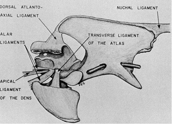
  • Instead of an intervertebral disc, 5 ligaments help stabilize the joint between C1 (atlas) and C2 (axis). The apical ligament, 2 alar ligaments, and transverse ligament are associated with the odontoid process (dens). The dorsal atlantoaxial ligament attaches the spinous process of the axis with the dorsal arch of the atlas. See Figure 3 below.
  • Congenital malformation of the dens (hypoplasia or aplasia) is common in toy breeds, which causes instability of the atlantoaxial joint and predisposes to subluxation. The most common presentation is dorsal subluxation of the axis (including the dens) causing ventral extradural compression of the cranial cervical spinal cord.
  • While young toy breeds are the most commonly impacted patients, it has been reported in older animals (like Milo) and larger breed dogs.
  • The degree of neurological dysfunction can vary greatly from neck pain alone to tetraplegia with respiratory compromise. When examining patients with suspected atlantoaxial instability extreme caution should be used when manipulating the neck, ESPECIALLY VENTROFLEXION, to avoid the peracute exacerbation of neurological signs and in some cases mortality.
  • An MRI is recommended for diagnosis to view the integrity of the spinal cord and associated ligaments, however cautiously acquired cervical spine radiographs can also be used in some cases for a definitive diagnosis.
  • Cervical bandaging with strict cage rest is recommended for a minimum of 8 weeks, along with analgesic medications and a corticosteroid taper. In some cases, surgical stabilization may be recommended (following MRI and CT imaging for planning).
  • The prognosis for patients managed with splints who have mild to moderate neurological deficits has been reported as fair to good. Depending on the severity of clinical signs, the diagnosis of this condition should not automatically lead to humane euthanasia if owners are open to serial bandaging and rest. The current treatment of choice for the majority of patients with atlantoaxial subluxation is splinting rather than surgical intervention.
  • Landry and Dr. Eifler are happy to consult on neurological cases by phone at 207-878-3121 or email specialty@pvesc.com.

Figure 3. Anatomy of the atlantoaxial joint and supporting ligaments.

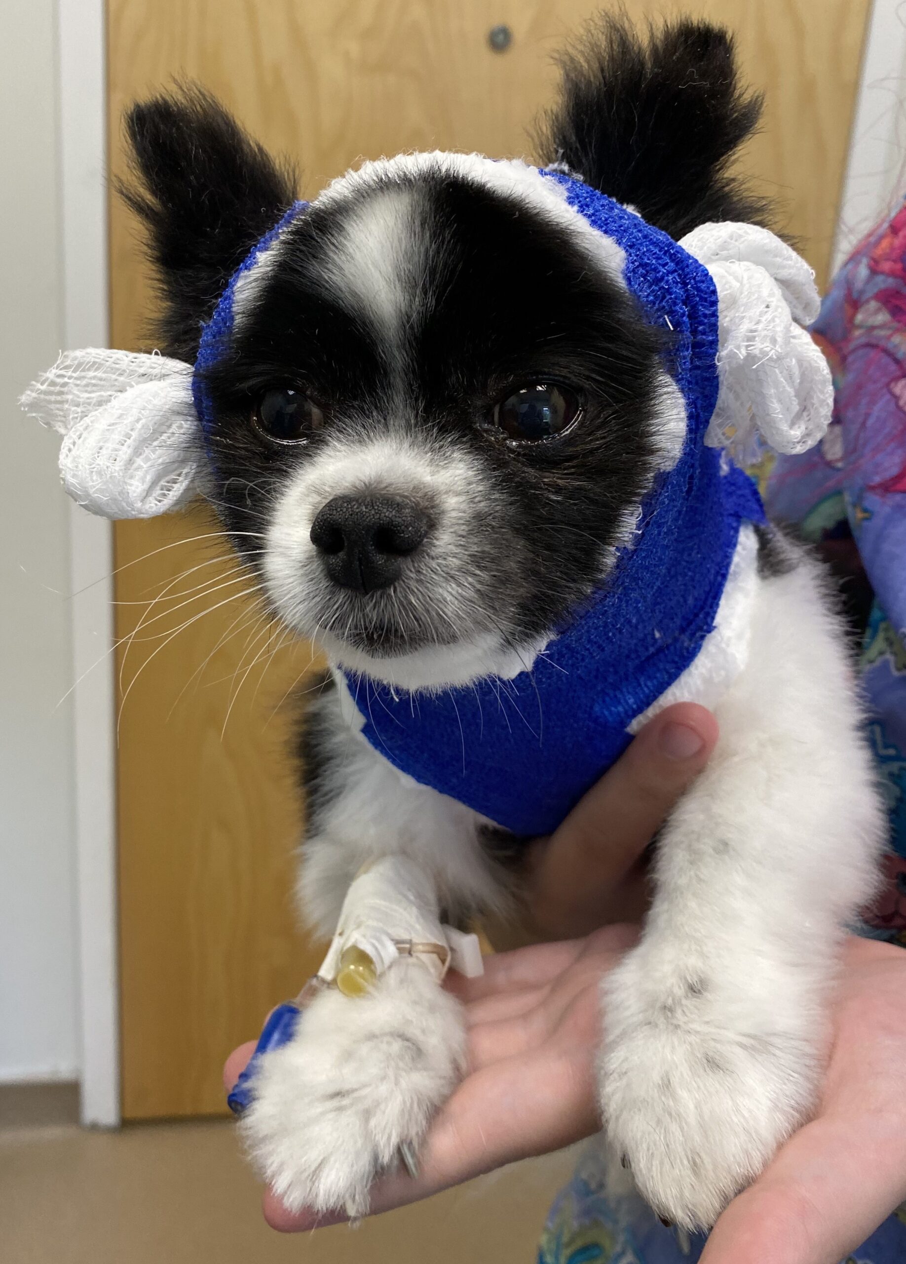
Figure 4. Milo with his first cervical stabilization bandage.

Sources:

Dewey, Curtis W., and Ronaldo C. Da Costa. Practical Guide to Canine and Feline Neurology. Third edition. Wiley Blackwell, (2016): 361-62.

Gage, E D, and J E Smallwood. “Surgical repair of atlanto-axial subluxation in a dog.” Veterinary medicine, small animal clinician : VM, SAC vol. 65,6 (1970): 583-92.

Authored by: Amanda Landry, DVM, DACVIM (Neurology)


Lucy is a 12 year old spayed female mixed breed dog that presented to the PVESC Internal Medicine Service for additional evaluation of a left adrenal mass.

Lucy developed clinical signs, including an increase in thirst and urination, in October 2023. Initial diagnostics were performed by the referring veterinarian. Lucy’s chemistry profile showed a mixed elevation in her liver enzymes (ALT 235, ALP 296, GGT 26). A complete blood count was unremarkable. Lucy’s total T4 was within normal limits. A urinalysis showed dilute urine without evidence of infection or inflammation. She later had her blood pressure measured. In January 2024, prior to starting anti-hypertensive therapy, Lucy’s blood pressure was found to be markedly elevated (> 200 mmHg). Enalapril was started as an anti-hypertensive. Lucy then had an abdominal ultrasound with her primary veterinarian that showed a cranial pole left adrenal mass. She was referred at this time.

Lucy first visited PVESC in March 2024. A recheck chemistry profile showed progressive elevations in her liver values (ALP 672, ALT 260, GGT 51). Dr. Shoelson, one of the PVESC internists, repeated Lucy’s ultrasound. A left, cranial pole adrenal mass that measured up to 2.94cm in diameter was appreciated. There was no evidence of vascular invasion. A sonographic image of the left adrenal mass is provided below.

Lucy had mild enlargement of her liver with some hyperechoic nodules distributed throughout. Hyperechoic nodules (likely myelolipomas) were noted in the spleen as well.

Adrenal tumors can be functional/hormone-secreting or non-functional. They can also be benign or malignant. An adrenal tumor that measures greater than 2 cm in diameter or that is invading into local vasculature is more likely to be malignant. The two most common malignant adrenal tumors are adrenocortical carcinomas and pheochromocytomas (which arise from the adrenal medulla and produce excess catecholamines). Similar clinical signs and biochemical changes can be seen with functional adrenocortical carcinomas and pheochromocytomas. Given Lucy’s cholestatic liver enzyme elevation and her increased thirst and urination, both classic changes seen with hyperadrenocorticism, we first elected to perform an ACTH stimulation test. Lucy’s ACTH stimulation test supported a diagnosis of hyperadrenocorticism. Our index of suspicion for a functional, adrenocortical tumor increased.

Lucy’s owner expressed an interest in moving forward with surgery. To stabilize her prior to surgery, we continued with anti-hypertensive therapy (enalapril) and also began treating her hyperadrenocorticism with twice daily trilostane (Vetoryl). Lucy’s clinical signs improved. A recheck chemistry profile showed improved liver enzyme values, and a follow up ACTH stimulation test was suggestive of ideal hyperadrenocorticism control. Lucy was then referred to Dr. Maxwell Bush to discuss minimally invasive laparoscopic adrenalectomy. Additional information on Lucy’s surgical treatment can be found in this Surgery article.

Authored by: Adam Shoelson, DVM, DACVIM (Small Animal Internal Medicine)


Rolly is a 4-year-old SF Boston Terrier who presented to our Neurology service for progressive difficulty walking in her forelimbs.

Case Summary:

Rolly presented for a 2-month history of neck pain and weakness in her front left limb, which had significantly progressed over the past several days to her being unable to walk on her own. Oral gabapentin and meloxicam only minimally improved her signs at home.

Neurology examination:

  • During her emergency neurology consultation, Rolly was unable to walk on her own due to significant left hemiparesis (worse in the left forelimb) and significant neck pain.
  • She had decreased to absent proprioception in the left forelimb and both pelvic limbs.
  • Her withdrawal and spinal reflexes (trigeminal and bicipital) in the left forelimb were significantly decreased.
  • Her neurological examination was most consistent with a C6-T2 myelopathy (worse on the left) and she was admitted for an emergency cervical MRI due to the progressive and severe nature of her signs.

MRI findings (see Figure 1 below):

  • Cervical MRI revealed a markedly hyperintense swelling associated with the spinal cord from C2 – T1 with loss of CSF patency but no extradural spinal cord compression. The intervertebral discs were all well-hydrated. There was severe contrast enhancement of the dorsal aspect of the spinal cord with suspected meningeal enhancement.
  • This MRI finding is consistent with severe meningomyelitis (inflammation of the cervical spinal cord parenchyma and meninges).
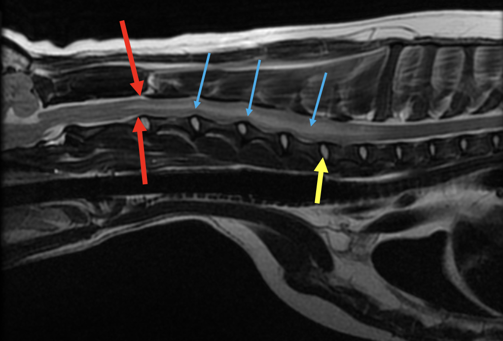
Figure 1. T2 weighted sagittal (midline) view of the cervical spinal cord

The cervical spinal cord parenchyma is markedly hyperintense which is shown by the blue arrows. There is significant spinal cord swelling present, as there is loss of CSF signal (shown cranial to the spinal cord swelling by the red arrows). The yellow arrow shows a (normal) hydrated intervertebral disc.

Infectious CNS disease testing and a cisternal CSF tap were submitted due to the need to differentiate between an infectious cause (protozoal, tickborne, viral, fungal, etc), immune-mediated cause, and less likely neoplastic cause (lymphoma). She was sent home the next day on oral antibiotics that cover the most common causes of infectious CNS disease in Maine (clindamycin, doxycycline, and enrofloxacin) while her tests were pending. She was also given oral prednisone in case she neurologically worsened over the weekend despite antibiotic therapy.

CSF and infectious disease testing results (see Figure 2 below):

  • Rolly’s CSF cytology revealed a significant lymphocytic pleocytosis (430 nucleated cells/uL) and elevated protein (112.6 mg/dL).
  • Her infectious testing was positive for toxoplasmosis on IgG (1:25) and IgM (1:50).
  • She was negative for tickborne disease, cryptococcus, distemper (titer consistent with vaccination only), and Neospora.
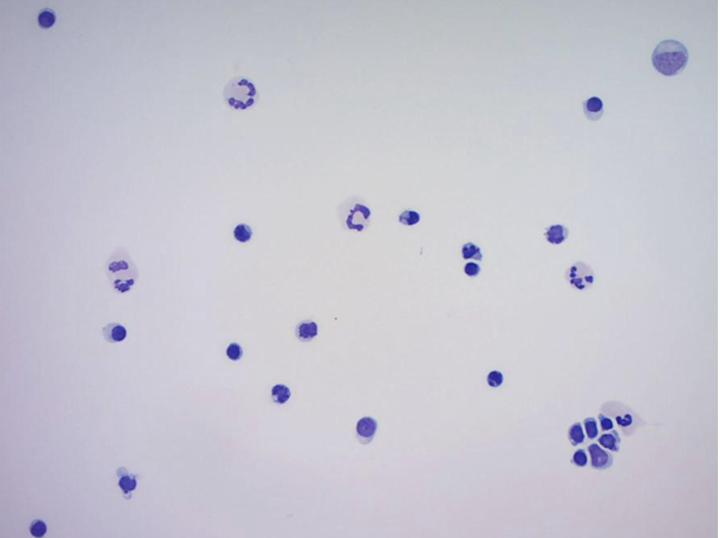
Figure 2. Cytospin slide prepared from submitted cisternal CSF sample.

The CSF sample consisted of 65% lymphoid cells, 24% neutrophils and 11% large mononuclear cells. The lymphoid cell population observed were small to intermediate in size and well differentiated. No infectious organisms or cellular atypia were observed.

Clindamycin was recommended for 6 weeks total and a recheck neurological examination was scheduled for 2 weeks-post imaging or sooner if needed.

Rolly rechecked with Dr. Landry as she continued to neurologically decompensate despite oral clindamycin therapy. At this time oral prednisone was started (0.8 mg/kg every 24 hours) and Rolly neurologically improved within a few days of starting corticosteroids. Due to a neurological plateau on this dose, her dose was increased to an immunosuppressive one (1.8 mg/kg daily) and she continued to improve and walk on her own.

Rolly did well on immunosuppressive prednisone but repeatedly worsened as her prednisone was tapered. Dr. Landry initiated injectable cytosine arabinoside therapy on an outpatient basis, and Rolly as of the publication of this article is neurologically normal on 0.9 mg/kg prednisone daily and injectable cytosine arabinoside every 4 weeks. She is 6 months out from her original diagnosis, and she is slowly being tapered off her medications.

The long-term goal in patients with immune-mediated inflammatory CNS disease is to gradually taper them off prednisone over months, and then eventually off injectable cytosine arabinoside (or other immunosuppressant medications).


Rolly has a mildly hypermetric gait in her forelimbs but has an otherwise normal neurological examination as of the date of this publication. We are so proud of (and happy for) her as she continues to live a great quality of life with her family!

Clinically relevant facts:

  • Immune-mediated meningoencephalomyelitis in dogs is a major differential for progressive neurological signs, especially in small to medium breeds.
  • In this author’s experience immune-mediated meningoencephalomyelitis does not carry the grave prognosis seen in previously published literature, and many patients can be weaned off medications completely (or continue a good quality of life on the lowest effective dose of injectable +/- oral medications). There is a population of these patients who are refractory to treatment and do not respond despite aggressive immunosuppression, or who cannot be weaned off high doses of immunosuppressive medications.
  • Cytosine arabinoside is an injectable anti-neoplastic medication that is used as an immunosuppressive agent in dogs. It is specific to the S phase of the cell cycle (DNA synthesis) and blocks cell progress from G1 phase to S phase.  This drug has excellent penetration into the CNS making it a great option for inflammatory CNS disease as well as CNS lymphoma. This author’s starting protocol includes one subcutaneous injection of cytosine every 24 hours for two doses. This protocol is starts at an every 3-week interval and is then slowly pushed out as the oral prednisone is tapered. It is generally very well tolerated at the dose used for inflammatory CNS disease. Adverse side effects include myelosuppression, gastrointestinal upset, and liver toxicity at high doses.
  • PVESC has an active out-patient cytosine arabinoside program for treatment of inflammatory CNS disease. This includes immune-medicated meningoencephalomyelitis, steroid-responsive meningitis arteritis (SRMA), and some other immune-mediated CNS diseases. Please do not hesitate to reach out to Dr. Landry or Dr. Eifler with any questions regarding this program (at specialty@pvesc.com).

Authored by: Amanda Landry, DVM, DACVIM, Neurology


Lucy is a 12 year old spayed female mixed breed dog that presented to Dr. Maxwell Bush of the PVESC Surgery Department for treatment of a left-sided adrenal mass. She was previously evaluated by Dr. Adam Shoelson of the PVESC Internal Medicine Department. Dr. Shoelson diagnosed with Lucy with hyperadrenocorticism, likely secondary to her adrenal tumor. Thus, our suspicion of index for a functional adrenocortical tumor increased. Additional information on the pre-operative management of Lucy’s hyperadrenocorticism can be found in the this Internal Medicine article.

With medical management resulting in ideal hyperadrenocorticism and improved blood pressure control, Lucy was deemed a good candidate for anesthesia and surgery. Furthermore, her medium to large size, the left-sided nature of her adrenal mass (the left adrenal gland is surgically more accessible), and the lack of obvious metastasis or vascular invasion, allowed us to recommend a laparoscopic approach for her adrenalectomy. Laparoscopic surgery, both in people and animals, has been shown to reduce peri-operative morbidity and mortality significantly. A video from Lucy’s laparoscopic procedure is provided below.


Lucy’s left adrenal tumor was successfully excised laparoscopically. She had a very stable anesthesia and recovery. She was discharged just one day after surgery. Trilostane (Vetoryl) and enalapril therapy were discontinued. A physiologic daily prednisone dose was administered until her first Internal Medicine recheck appointment. Lucy’s adrenal mass was submitted for histopathology, which was consistent with a narrowly excised adrenocortical carcinoma without evidence of vascular invasion.

Through Lucy’s post-operative rechecks with Dr. Shoelson, her liver enzymes improved and then normalized. She is now non-hypertensive. Lucy is clinically normal at home.

Three weeks post-operative, Lucy’s owners sent us a fantastic update: “She is like a puppy.  She runs all over, eats her meals and treats and drinks her water normally, not in excess. Her coat is finally shedding after almost two years and it looks so full and healthy.  It had been so dry, dark, and oily-looking, with no shedding!  This was unheard of for a dog with Pyrenees in her but no one seemed to think it strange except us. Thank you so much.  It was amazing that the surgery could be done laparoscopically by Dr. Bush and thankfully  was recommended by Dr. Shoelson. We are so happy that Lucy had such a wonderful place to go to for medical services.  You are all so kind, caring and very knowledgeable.”

For the future, we would like to continue to pursue minimally invasive laparoscopic surgeries for a variety of procedures, including adrenalectomies and cholecystectomies. However, ideal patient selection can be challenging and is of paramount importance to ensure success.

Authored by: Maxwell Bush, VMD, DACVS-SA, Surgery


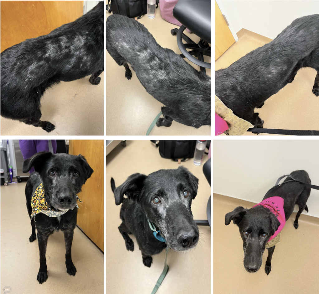
Lucy was presented to our Oncology service for consultation following a diagnosis of cutaneous epitheliotropic T-cell lymphoma (CETL) via skin biopsies performed by her primary care veterinarian. In early August 2022, Lucy was presented to her primary care veterinarian for hair loss and seborrhea. Fungal culture yielded Microsporum vanbreuseghemii and bloodwork, which included T4, was unremarkable. Treatment with Atopica was initiated with minimal response. Lucy returned to her primary care veterinarian at the end of August 2022 for weight loss, at which time a skin scrape was negative for Demodex. She was treated with an injection of ivermectin and prescribed a medicated shampoo, oral antibiotics and steroids. Lucy’s cutaneous lesions persisted, so biopsies of the affected areas were performed in late September 2022, yielding a diagnosis of CETL. Referral to oncology was recommended at that time. 

On examination at her initial consultation in November 2022, Lucy had diffuse non-erythematous scaling with multifocal to coalescing alopecia along her dorsum (see photo). Patchy alopecia and seborrhea was present along her face and all four limbs. She had a couple subcutaneous masses that were presumed to be lipomas and her lymph nodes palpated within normal limits. Further staging with chest X-rays and an abdominal ultrasound were recommended but were declined at that time. 

The gold standard treatment for patients with diffuse CETL is a combination of prednisone and chemotherapy. The most commonly used first-line drug is an oral chemotherapy agent called lomustine (CCNU) administered once every 3 weeks until cancer progression or toxicity occurs. The median duration of remission reported for dogs with diffuse CETL and treated with lomustine is about 6 months. Since cancer cells in CETL are primarily confined to the skin, chemotherapy agents must be lipophilic in order to accumulate within the skin and be effective. Alternative therapies used in the management of CETL include oral retinoids (isotretinoin), Apoquel (anecdotal evidence of responses), Tanovea (injectable), and radiation therapy, among others. Lomustine rarely causes gastrointestinal upset but often causes marked bone marrow suppression (neutrophils and platelets) and liver enzyme elevations. 

The owners elected to initiate treatment with lomustine and Lucy received her first treatment at the time of her initial consultation in November 2022. Lucy experienced a grade 3 non-febrile neutropenia 1 week after her first treatment and a grade 1 non-febrile neutropenia 3 weeks after her first treatment. Prophylactic treatment with Baytril was initiated and the frequency of her treatments was decreased to every 4 weeks to give her bone marrow more time to recover. The scaling over Lucy’s dorsum worsened after her first couple treatments, which is often observed in patients responding favorably to treatment as the affected skin begins to flake off. Lucy’s cutaneous lesions slowly resolved with each subsequent treatment (see photos). 

Photographs of Lucy showing resolution of cutaneous lesions over the course of her chemotherapy treatment. Diffuse cutaneous lesions were observed at initial presentation on November 2, 2022 (LEFT). Following her first lomustine treatment, Lucy’s seborrhea and scaling appeared transiently progressive on November 9, 2022 (MIDDLE) before improving. Significant improvement was observed at the time of her second treatment, on December 7, 2022 (RIGHT). 

Lucy appears to be in complete remission in February 2023 (LEFT) and June 2023 (RIGHT). 

Treatment was discontinued after 6 doses due to persistent neutropenia that worsened after each subsequent dose. Alternative treatment options were discussed and the owners elected to transition her to monitoring. Lucy remained in remission until she was humanely euthanized due to mobility issues in April 2024. She was truly a cancer survivor and her story serves as a reminder that pets can have a great quality of life while undergoing chemotherapy and treatment may result in an unexpected favorable outcome! 

Clinically Relevant Facts:

  • The prognosis for cutaneous epitheliotropic T-cell lymphoma (CETL) varies depending on the extent of disease with diffusely affected patients experiencing short survival times. 
  • Treatment involves chemotherapy and lomustine (CCNU) is most often used as first-line therapy due to lipophilic properties that allow it to concentrate within the skin. 
  • The majority of patients experience cancer progression after 3-4 doses and alternative therapies, such as retinoids (isotretinoin), Tanovea, Apoquel (anecdotal evidence) and radiation therapy, among others, may be considered for continued management. 
  • Chemotherapy is generally well-tolerated and durable complete responses have been observed in some patients.

Authored by: Brittanie Partridge, DVM, PhD, DACVIM, Oncology


Elvis was a 5 yr MN Great Dane who presented to our surgery service for a mass at the level of the right caudal chest. His owners reported that they first noticed the mass about three months ago and it had been rapidly growing since then. Besides the mass he appeared comfortable and was otherwise eating and drinking well.

Dr. Marta Agrodnia performed a thoracic CT which revealed a large contrast enhancing mass along the right rib cage, with growth into the thoracic space. A surgical biopsy was obtained under general anesthesia, which returned as favoring a chondrosarcoma. The pathologist noted 4 mitoses in 10 high power fields (MI = 4). There was no vascular invasion noted throughout the biopsy and the pathologist suggested the microscopic features were most suggestive of a grade I chondrosarcoma.

Elvis’s surgical biopsy following thoracic CT: The poorly demarcated and highly cellular mass is composed of polygonal cells embedded in lacunae which are separated by abundant chondroid matrix. Multifocally, there are 2-4 cells per lacunae with neoplastic cells having moderate nuclear pleomorphism.

Surgical resection was recommended due to the invasive nature of the mass and increased survival time associated with tumor excision. Due to the rapid rate of growth it was discussed with his owners that there was a risk of a more guarded prognosis, if excisional biopsy revealed evidence of osteosarcoma.

Both mass removal and rib resection were performed by Dr. Agrodnia (pictures included at the bottom of this article):

  • Preoperative ultrasound-guided intercostal blocks were performed by Dr. Raphael Vizena using ropivacaine
  • An elliptical incision was made over the right caudal thorax. Hemostasis was managed with both electrocautery and Ligasure.
  • All visually abnormal tissue was removed, along with 2-4 cm margins on all normal appearing fat and muscle.
  • A sagittal saw was used for resection of ribs 8, 9, 10, and 11
  • The diaphragm was reattached to the body wall using interrupted cruciate sutures
  • Two pieces of SurgiMesh (15 x 7.5 cm) were placed in the chest wall defect and then secured into place with interrupted cruciate sutures, taking care to keep the knots external to the mesh and buried into tissue.
  • The two mesh pieces were also secured together, and a chest tube was placed under mesh.
  • Closure was performed in two layers and Nocita was placed locally
  • A trans-costal epidural soaker catheter was placed as well as a sterile bandage.

Elvis recovered uneventfully from surgery. The mass Dr. Agrodnia excised weighed 3.06 kg, and histopathology confirmed the diagnosis of a chondrosarcoma. As of the date of this publication, Elvis has had no recurrence of his rib mass. He met with our oncology service to initiate metronomic chemotherapy (chlorambucil) shortly following surgery, to decrease the risk of tumor regrowth.

CLINICALLY RELEVANT FACTS:

  • Chondrosarcoma is the second most common primary bone neoplasia in canines. Dogs who are medium to large breed, and middle age to older are predisposed. These tumors arise from cartilage and tend to involve flat bones (ribs, turbinates, pelvis, etc). They are considered malignant tumors, however they often have a low metastatic rate with some data suggesting a rate of 20% or lower.
  • Surgical excision is recommended to decrease the risk of metastasis, and therefore prognosis has historically been linked with anatomical location (depending on the potential for complete excision). Local invasion and recurrence can occur after surgery, so owners should be warned of these risks.
  • The major differential for chondrosarcoma includes osteosarcoma, which carries a much higher metastatic rate and guarded prognosis. Caution should be used when discussing prognosis and making a treatment plan in cases like Elvis, where a small surgical biopsy (in comparison to the entire 3.06 kg tumor) is used to diagnose the tumor type. Luckily Elvis’ surgical excision confirmed chondrosarcoma, however surgical biopsy in similar cases may underestimate the grade or misdiagnose the tumor type depending on which part of the mass was sampled. If excisional biopsy was suggestive of osteosarcoma, a more guarded prognosis for metastasis would have been reported to the owner. This would also impact how the PVESC oncology service would manage his case.

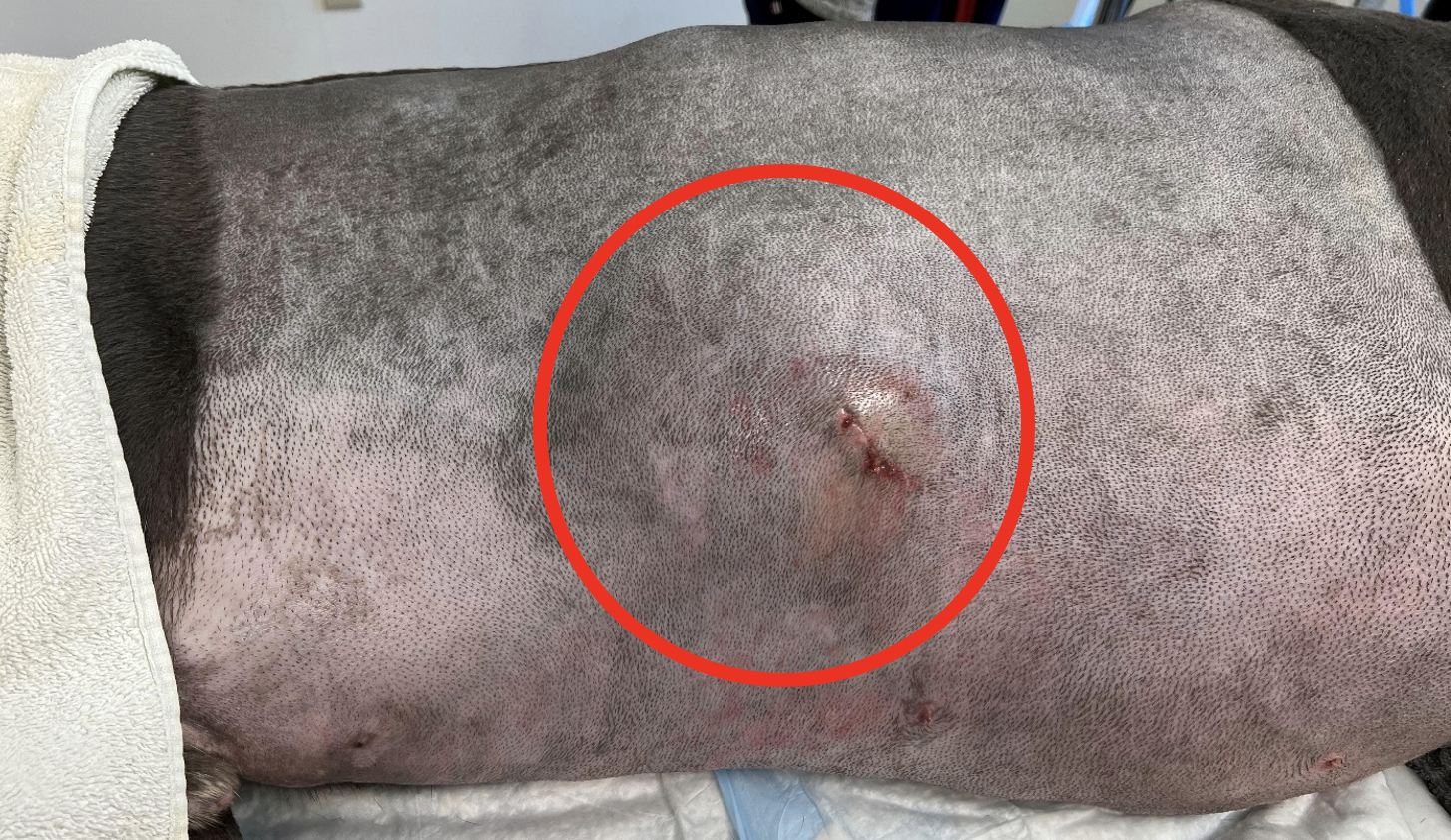
The red circle outlines the border of the right rib mass, visible just prior to surgery. This mass had been visible to the owners for about 3 months and they felt it was rapidly growing and non-painful. The previous incisional biopsy site (post thoracic CT) is visible.             

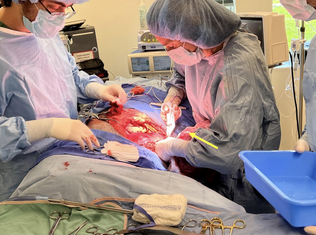
Dr. Agrodnia has the large tumor in her arms (the tumor can be seen labeled by the yellow arrow), carefully removing the last few connections between the tumor and Elvis’ body.

The tumor weighed 3.06 kg upon removal.

Both pieces of mesh are sutured into the chest wall defect (white arrows), and to each other (purple arrow). The chest tube can be seen below the mesh.

Authored by: Dr. Landry, DVM, DACVIM, Neurology 


Junior was presented to our Oncology service for consultation following a diagnosis of cutaneous hemangiosarcoma made via biopsy of a mass removed by his primary care veterinarian. The mass had been present for about 4 months and started to grow rapidly leading up to its removal. Histopathology of the mass was most consistent with a diagnosis of cutaneous hemangiosarcoma. Unfortunately, invasion into subcutaneous tissue was observed and complete excision could not be confirmed. Both variables have been associated with an unfavorable prognosis.

On examination, Junior had a few other cutaneous and subcutaneous masses, one of which had some bruising resembling a second cutaneous hemangiosarcoma. Junior was staged completely with chest X-rays and an abdominal ultrasound, which did not reveal any evidence of metastasis. Since Junior was at a higher risk for local tumor recurrence and metastasis, treatment with chemotherapy was indicated in his case. Treatment options offered to the client included additional surgery to obtain wider margins, injectable doxorubicin therapy and/or metronomic chemotherapy, which involves continuous administration of low-dose oral chemotherapy.

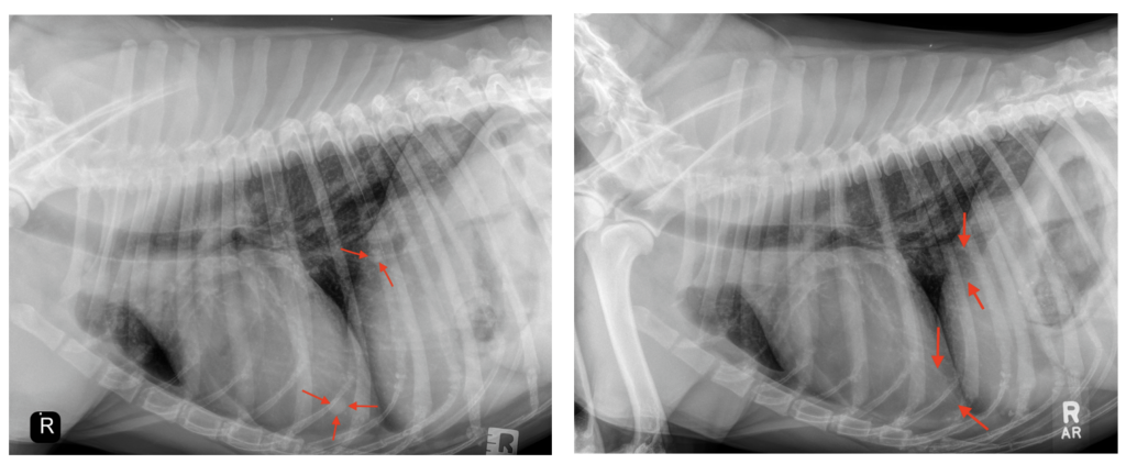
The owner elected to move forward with metronomic chemotherapy as this option has been associated with fewer side effects. Approximately 5 months after initiating therapy, Junior’s cancer progressed in the form of multiple new cutaneous masses and pulmonary metastasis (Image). Vorinostat, an alternative oral chemotherapy drug, was added to Junior’s treatment regimen at that time due to apparent synergistic antiangiogenic effects with metronomic chemotherapy. Vorinostat is histone deacetylase (HDAC) inhibitor, which allows for activation of genes involved in cell cycle arrest resulting in tumor cell death.

Thoracic radiographs taken in April (LEFT) revealed evidence of soft tissue nodules (arrows) consistent with pulmonary metastasis. Repeat radiographs performed in July (RIGHT) to assess Junior’s response to combination therapy revealed complete resolution of the previously noted nodules (arrows).

Approximately 3 months after initiating combination therapy, Junior presented to PVESC for restaging to assess his response to treatment. On examination, ALL of Junior’s previously noted cutaneous and subcutaneous masses had completely resolved and chest X-rays revealed complete resolution of all pulmonary nodules (Image). Continued treatment with combination therapy until either cancer progression or toxicity was recommended at that time. The PVESC Oncology team feels honored to have been a part in Junior’s cancer journey.

CLINICALLY RELEVANT FACTS:

  • Cutaneous hemangiosarcoma typically carries an excellent prognosis following complete surgical excision.
  • Invasion into the subcutaneous tissue and incomplete excision may significantly impact overall prognosis as in Junior’s case. Thus, complete tumor removal with wide margins is vital to improving long-term outcome.
  • Novel approaches to cancer management with targeted therapies, such as Vorinostat, have improved our ability to successfully manage cancer patients with advanced disease.

Authored by:  Dr. Brittanie Partridge, DMV, PhD, DACVIM, Oncology


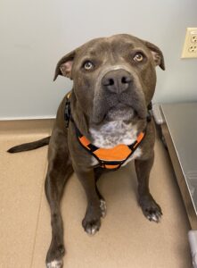
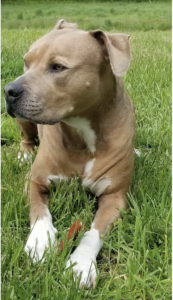
Opal is a 1.5 year SF Pit Bill Mix who presented to our Neurology service for a one-week history of dragging her pelvic limbs intermittently. Opal first started showing symptoms after running in the woods with her housemates. That evening she began limping in her pelvic limbs, and since then Opal had been only able to walk about 30 feet before she would start to drag her pelvic limbs behind her. Her owners report that with a period of rest she is able to get up in her back legs to attempt to walk again. Over the past few days she developed diarrhea and has not been eating well. A CBC, 4DX, and radiographs of the abdomen/thoracolumbar spine were unremarkable at the emergency veterinary hospital. Due to concern for back pain she was started in carprofen, methocarbamol, metronidazole, and gabapentin which did not improve her signs.

On neurological examination, Opal was bright and alert but nervous. When she rose from rest, she would walk 5-6 steps before she became progressively weak in the pelvic limbs, bunny hopped, and then collapsed in the forelimbs as well. After a minute of rest she would repeat this gait, until she collapses in all four limbs. Videos have been provided below. Cranial nerve examination was unremarkable, except for a fatiguing palpebral reflex. Her conscious proprioception was unremarkable in all four limbs, as were all her spinal reflexes. No pain was found on cervical range of motion or deep thoracolumbar and sacral spine palpation. Dr. Landry neuro-localized her to the neuromuscular junction.  At her appointment thoracic radiographs, CBC/chemistry, and a T4 were unremarkable. Abdominal ultrasound was consistent with her recent colitis but was otherwise unremarkable. Due to the concern for myasthenia gravis, an acetylcholine receptor antibody titer (AChRAb titer) was submitted and Opal was started on pyridostigmine bromide.

This first video shows Opal’s gait after allowing her to rest in sternal recumbency for several minutes.


This second video shows Opal after we asked her to continue to walk following rest. The progression of pelvic limb weakness over multiple steps to bunny hopping and then collapse is typical of myasthenia gravis in dogs.


Her owners reported that Opal’s neurological signs improved after several doses of pyridostigmine bromide. Her dose was titrated up to the lowest effective dose that keep her neurologically improved. Her AChRAb titer confirmed the diagnosis coming back as 1.82 nmol/L (normal serum titer <0.6 nmol/L).  As of the publication of the article, Opal was doing well at home on pyridostigmine bromide therapy alone.

CLINICALLY RELEVANT FACTS:

  • Acquired myasthenia gravis is an autoimmune condition in which there are autoantibodies made to the acetylcholine receptors at the neuromuscular junction. In some cases this may be paraneoplastic (secondary to a thymoma or other type of cancer), so thoracic radiographs and abdominal ultrasound are recommended. Dogs with paraneoplastic myasthenia gravis will not resolve spontaneously, and referral for removal of the tumor is often recommended. Thoracic radiographs are also recommended in order to screen for megaesophagus, as this is considered a common condition in dogs with myasthenia gravis which require elevated feedings and sometimes medications in order to avoid regurgitation and aspiration pneumonia.
  • There are a small percent (previously reported to be 2%) of myasthenic dogs that are seronegative (have a negative or low titer). False positives are extremely rare. These dogs should be retested one month later, as some of them will convert and have a positive titer on recheck. There are several theories regarding seronegative myasthenia gravis, most common being that some dogs have autoantibodies against receptors on the NMJ end plate OTHER THAN acetylcholine (like titin or ryanodine receptors).
  • Pyridostigmine bromide is an “anti-acetylcholinesterase” medication which, by inhibiting the acetylcholinesterase enzyme from breaking down acetylcholine, increases the bioavailability of acetylcholine and therefore increases the ability of nerve impulse transmission at the neuromuscular junction. This will clinically improve dogs with myasthenia gravis, but dogs with other neuromuscular junctionopathies can also improve clinically. Therefore a AChRAb titer should be submitted regardless of clinical improvement.
  • Treatment should be continued until there is seroconversion of the AChRAb titer. The high risk of aspiration pneumonia has lead to the previously reported 50% one year mortality rate. However spontaneous remission has been reported in 80% of dogs within 6-8 months of diagnosis.
  • Some cases will fail pyridostigmine bromide medication alone, and require the additional of immunosuppressive medications.
  • Besides the hallmark appearance of their gait, dogs with myasthenia gravis will often have normal spinal reflexes and a fatiguing palpebral reflex.


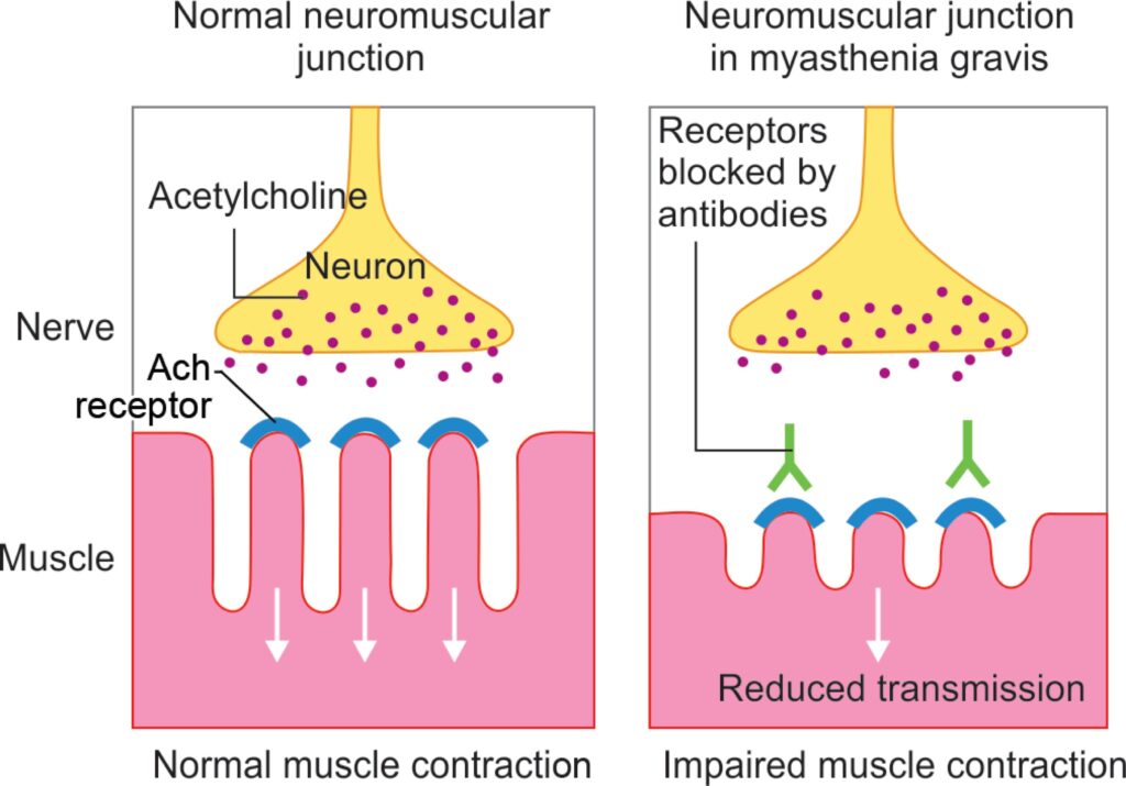
The normal neuromuscular junction vs the neuromuscular junction in a canine with myasthenia gravis. The patient with myasthenia gravis has acetylcholine receptors which are unusable as they are bound by autoantibody. These patients have limited neuromuscular transmission at these junctions and are clinically weak with exercise.

Authored by: Dr. Landry, DVM, DACVIM, Neurology 


Willow is a 3 year old FI Lab Mix who was admitted to our emergency service after being trapped in a housefire. She was found by a Portland firefighter (from the Munjoy Hill Fire Station) who administer oxygen on site prior to transportation. On presentation she was dyspneic, covered in soot, and had harsh lung sounds on thoracic auscultation.

Intranasal catheters were placed for oxygen administration. Bloodwork showed hypophosphatemia (1.6 mg/dL – suspect secondary to hyperventilation), a mild elevation in ALT (356 U/L), and GGT (16 U/L). Both cornea showed evidence of superficial corneal ulceration, and Willow was coughing up black soot material.

Her owners opted to admit Willow on high-flow oxygen via the intranasal catheters. During her hospitalization Willow also received IV sedation (as needed), IV unasyn, IV fluids, and albuterol inhalant treatment. Willow was hospitalized for a total of 7 days, 6 of which were on high flow oxygen.

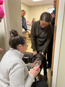
After a long 6 days of fighting, Willow was discharged to her owners having been off of high flow oxygen for 24 hours. Her owners and the Portland firefighters involved in her rescue arrived to our Warren Ave location to cheer her on as she was discharged from the PVESC Emergency/Critical Care service. As of this publication Willow has continued to improve at home, and we are so happy for her continued recovery.

Clinically relevant facts:

  • The pathophysiology of smoke inhalation is multifactorial. The progression of clinical signs and prognosis differ depending on present respiratory irritants, thermal heat exposure, particulate matter size, and systemic toxins.
  • Thermal heat damage more commonly impacts the upper airway mucosa, leaving dogs vulnerable to upper respiratory infections (chronically). Dogs with prolonged/high heat exposure causing lower airway thermal damage carry a more guarded prognosis as they are prone to edema-driven upper airway obstruction.
  • Carbon monoxide exacerbates the clinical signs, if present, in smoke inhalation cases. It is rapidly absorbed across the alveolar membrane and has 200-250 x greater affinity for hemoglobin than oxygen. This left shift of the oxygen-hemoglobin dissociation curve causes a secondary cellular hypoxia. Administration of oxygen by first responders is imperative to accelerate the elimination of carbon monoxide and support arterial oxygenation.
  • High flow nasal oxygen delivers more concentrated oxygen than traditional oxygen, and allows many patients to avoid the need for mechanical ventilation.

Authored by: Dr. O’Brien, DVM, DACVECC, Emergency and Dr. Landry, DVM, DACVIM, Neurology


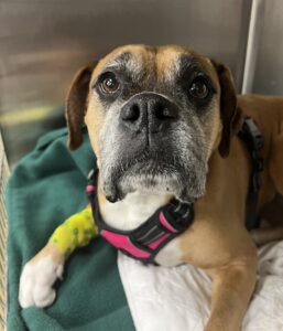
Pearl is a 6.5 yr old spayed female Boxer that was transferred to our Internal Medicine Service (Dr. Adam Shoelson DVM, DACVIM) after being admitted to the hospital through the Emergency Service for severe, unilateral epistaxis and collapse. Chronic nasal signs (purulent-appearing nasal discharge, epistaxis, sneezing) had previously been longstanding in nature. Five months earlier she had been diagnosed with and treated for chronic bacterial rhinitis after an extensive, advanced diagnostic work-up performed by another specialty clinic. She failed to improve with this rhinitis therapy. Her signs appeared to be refractory to antimicrobial therapy. 

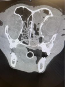
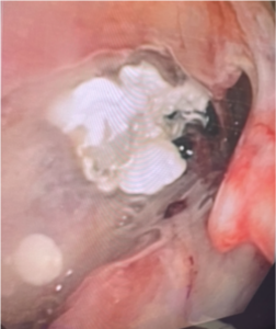
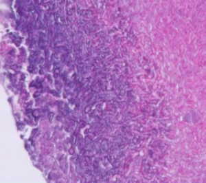
Previously visualized destructive nasal changes noted on her initial CT scan raised doubt about her chronic rhinitis diagnosis. It was decided that advanced diagnostics should be repeated. Pearl received a packed red blood cell transfusion due to clinically significant anemia that had developed secondary to her epistaxis. A CT scan of Pearl’s head showed severe destructive changes to the left nasal cavity. Fluid or material was noted in her left frontal sinus (Figure 1). Rhinoscopy confirmed the destructive changes and also permitted visualization of fairly extensive fungal plaques in the nasal cavity and frontal sinus (Figure 2). Histopathology from nasal biopsies revealed intralesional fungal hyphal mats and pleocellular inflammation (Figure 3). Aspergillus was confirmed on fungal culture and antibody testing (aspergillosis immunodiffusion). Fungal debridement was performed to remove visualized plaques (Figure 4). An initial dose of topical, 1% intranasal clotrimazole cream was instilled. Pearl was started on oral itraconazole (5 mg/kg PO once daily). 

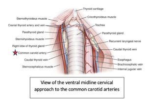
Unfortunately, Pearl represented for severe epistaxis and collapse several days later. A second packed red blood cell transfusion was administered. Due to concerns for intractable epistaxis caused by fungal damage to local vasculature, surgical left carotid artery ligation was performed. The Surgery Article of the Month can be referenced for additional information on this procedure. 

Pearl’s epistaxis resolved following her carotid artery ligation. This procedure allowed the Internal Medicine Service to move forward, freely, with numerous subsequent rhinoscopic evaluations and clotrimazole infusions. Given Pearl’s extensive fungal load, seven rhinoscopic debridement procedures/clotrimazole infusions were performed in total. Pearl has remained on itraconazole since her diagnosis. Her most recent rhinoscopy was performed in October 2023. No fungal plaques were appreciated, though Pearl did still have severe nasal turbinate destruction. Furthermore, light pink, friable polyps were noted in the left nasal cavity (Figure 5). Biopsy of these structures confirmed the presence of inflammatory polyps, changes likely being driven by inflammation caused by her previous fungal infection. A repeat fungal culture was negative. As Pearl has tolerated her itraconazole from a biochemical perspective (diligent monitoring of her liver enzymes has been performed), we have elected to continue this indefinitely over concerns for possible fungal disease relapse. 

Her owner reports she is feeling great at home and is loving life as of this publication date.

Clinically relevant facts:

  • Fungal rhinitis, though often a diagnostic challenge, should be considered as a differential for chronic sneezing, nasal discharge, and epistaxis in Maine, especially if destructive changes and frontal sinus material are noted with CT/rhinoscopy. The diagnosis of sinonasal Aspergillosis should, ideally, be confirmed with fungal culture or antibody testing (Aspergillus antibody immunodiffusion). 
  • As a rule of thumb, unilateral epistaxis is generally more concerning for focal disease (neoplasia, fungal disease, nasal foreign material). Bilateral epistaxis/discharge is concerning for more generalized disease like chronic rhinitis. Characteristic destructive CT changes should increase the index of suspicion for fungal disease or neoplasia dramatically. 
  • Sinonasal Aspergillosis treatment typically involves a combination of fungal plaque debridement (facilitated by rhinoscopy and/or sinus trephination), topical therapy (clotrimazole), and administration of systemic antifungal medications (often in very long-term fashion). Treatment can become very expensive depending on the number of anesthetized procedures required and the type of oral antifungal recommended. A need for blood transfusions and/or carotid artery ligation further increases cost. 
  • Though generally unnecessary, carotid artery ligation should be considered as a life-saving surgical intervention for cases that present with intractable epistaxis. 
  • Generally speaking, the prognosis for localized sinonasal Aspergillosis is fair, though the treatment can be long, highly involved, and expensive. In cases where there is intracranial involvement or systemic disease, the prognosis is guarded to poor. 

Figure 1: A head CT scan demonstrating severe left-sided nasal turbinate destruction and material within the left frontal sinus.

Figure 2: Rhinoscopic evaluation of the left nasal cavity confirmed severe destructive rhinitis. White, soft fungal plaques were also seen.

Figure 3: Histopathology of biopsies obtained from the left nasal cavity demonstrated significant pleocellular inflammation and mats of fungal hyphae.

Figure 4: Multiple rhinoscopic procedures were performed to guide debridement of fungal plaques in a minimally invasive fashion. A brown fungal plaque removed from the nasal cavity is shown.

Figure 5: The most recent rhinoscopy, performed primarily for monitoring purposes, demonstrated a nasal cavity clear of fungal plaques. However, there were light pink, friable polyp-like lesions. These were biopsied with results consistent with inflammatory polyps.


Authored by: Dr. Shoelson, DVM, DACVIM, Internal Medicine and Dr. Landry, DVM, DACVIM, Neurology 


Pearl was a 6 year old SF Boxer transferred to our Internal Medicine (Dr. Adam Shoelson) service after being admitted for unilateral epistaxis and collapse. After a blood transfusion a nasal CT and rhinoscopy were performed, which provided the diagnosis of destructive fungal rhinitis (see the Internal Medicine article for more information on Pearl’s case).

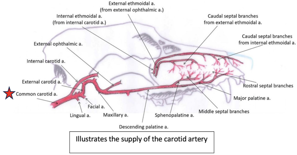
Pearl represented to our emergency service a few days after her procedure for another episode of anemia-induced collapse. The severity of the nasal vasculature destruction was causing life threatening hemorrhage. Dr. Gianetto from our surgery service performed a unilateral left carotid artery ligation under general anesthesia.

Since then, Pearl has continued treatment for her fungal rhinitis and has had no further collapse episodes. She has continued to do great as of the publication of this article under the care of our Internal Medicine service.

Clinically relevant facts:

  • Carotid artery ligation should be considered in dogs with clinically significant oral or maxillo-facial hemorrhage, where traditional hemostatic methods are unsuccessful or unfeasible.
  • Complications for this procedure in dogs are not well documented, but in general include retinal damage and cerebral edema.
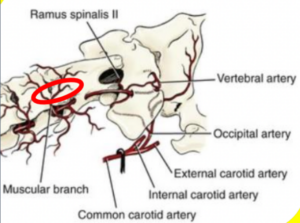
  • Anatomically dogs have sufficient arterio-arterial collateral blood supply from the vertebral arteries, and can therefore tolerate bilateral carotid artery ligation if needed.
  • Bilateral carotid artery ligation may predispose canine patients to strokes (ischemic events). No recommendations currently exist in veterinary medicine on whether anticoagulant therapies are indicated for ischemic event prevention.
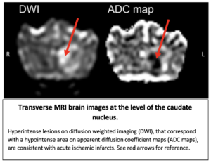
  • We have provided a case report for a 3 yr MI Border Collie who underwent life-saving bilateral carotid artery ligation to address uncontrollable oral and nasal hemorrhage. These signs occurred after an episode of stick chewing. 5 days post-surgery this patient presented for peracute onset forebrain signs, and was diagnosed with a striate artery infarct via MRI. See MRI pictures from this article.


Authored by: Dr. Landry, DVM, DACVIM, Neurology
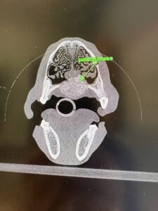
 

Mr. Smalls presented to our Neurology service for severe neck pain. He had episodes of crying out in pain and holding up his right front limb. Other than neck pain, he had no other neurological deficits on his examination at PVESC.

Cervical MRI revealed a severely lateralized foraminal disc herniation at C2-3 on the right. This was consistent with his clinical signs at home. Successful medical management was given a guarded prognosis due to the severe lateralization of the disc herniation.

Mr. Smalls was admitted for a cervical hemilaminectomy, as he had multiple recurrences of neck pain as oral prednisone was tapered. A cervical hemilaminectomy was performed by Dr. Eifler and Dr. Landry as a team, and Mr. Smalls was discharged several days later comfortable on oral medications for strict crate rest.

Mr. Smalls presented for his 6 week post-surgery recheck with a normal neurological examination off all oral medications.

The PVESC Neurology team is so happy for Mr. Smalls’ recovery, and are so thankful to have the dedicated team and quality facility to help pets with challenging neurological conditions.

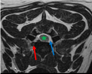
T2-Weighted transverse cervical spine MRI image. There is extradural nerve root compression on the right.

The red arrow shows the small round hypointense disc material compressing the right nerve root.

The left nerve root appears normal (blue arrow).

The spinal cord is labeled with a star.


CLINICALLY REVELANT FACTS:

  • Cervical disc herniations that involve the nerve root carry a more guarded prognosis for medical management alone (prednisone, rest, analgesic medications)
  • A routine ventral slot surgery has little to no ability to reach disc and nerve root in the foramina
  • Cervical hemilaminectomies require longer recovery (due to the dorsal approach) and carry higher risk due to the location of the vertebral artery
  • Red oval below shows ideal surgical site margins


ABOUT PVESC NEUROLOGY

PVESC Neurology is here for you Monday – Friday for scheduled appointments, and 7 days a week for neurological emergencies. Our state-of-the-art MRI allows us to offer the gold standard care for our patients 24/7. Our goal is to continue to bring quality veterinary neurology to the state of Maine.

If you are looking to refer and quote a client for emergency advanced imaging +/- surgery, please see general guidelines below:

Bloodwork, brain MRI, +/- CSF tap – $3500 – 4000. Infectious CNS disease testing may be recommended at additional cost pending test results.
Bloodwork, spine MRI, surgical decompression, hospitalization – $7000 – 10,000.

Authored by: Dr. Landry, DVM, DACVIM, Neurology
 

Portland Veterinary Emergency and Speciality Care (PVESC) has been serving Maine for over thirty years. At PVESC, we offer expert multi-specialty and emergency care services for our patients and peace of mind for our clients. We work in partnership with veterinary practices throughout New England to step in when more advanced care is needed.

We are proud to offer the services of Emergency and Critical Care (available 24/7/365), Surgery, Anesthesiology, Internal Medicine, Neurology, Oncology, Cardiology, Dermatology, Radioactive Iodine therapy for Hyperthyroid Cats, and Synovetin OA therapy for canine osteoarthritis.  

As an ever-growing, expanding, and evolving practice, we offer you the opportunity to continue to learn and grow with us. We foster a collaborative and supportive team culture, and we empower our teams to provide the best medicine possible for our patients. We understand the importance of work-life harmony and we encourage our team to take full advantage of all that Maine has to offer.

Core Values

At PVESC we believe in respect for the individual, the client, and the patient. We work together as a team and expect all members of that team to treat one another respectfully. Our goal is to provide the highest quality medical, surgical and critical care to our patients, and offer excellent service to our clients.


Benefits

PVESC is committed to valuing you by providing a rewarding compensation package that includes fair base pay commensurate with your experience, and an extensive benefit plan. Our benefits include:


Education and Advanced Training Opportunities at PVESC


Veterinary Student Externships:

PVESC hosts veterinary student externships and preceptorships in single or multiple disciplines, including Emergency, Internal Medicine, Oncology, Surgery, Ophthalmology, Dermatology, and Cardiology.  Please forward CV and letter of interest to Kristine at kristines@pvesc.com.

Veterinary Technician Student Externships:

PVESC hosts veterinary student externships in single or multiple disciplines, including Emergency, Internal Medicine, Oncology, Surgery, Ophthalmology, Dermatology, and Cardiology.  Please forward CV and letter of interest to Kim at kimk@pvesc.com.


PVESC is proud to support Pet Rock in the Park! Join us Sunday, August 25th, 2024, for this family-friendly event in Deering Oaks Park in Portland from 11 am – 3 pm. The stage will feature live music! Food and pet products will be available for purchase. Proceeds support the Animal Cancer Foundation. The event is pet friendly and FREE! Learn more about vendor opportunities here.

Special thanks to Nothing Bundt Cake in Scarborough, Monte’s Fine Foods, Coffee By Design, and Stephen King for their generous donations to our raffle for Pet Rock in the Park 2024, helping to support the fight against cancer.


PVESC’s next Continuing Education dinner event is scheduled for November 11, 2024, at the Holiday Inn by the Bay in Portland, Maine. Any questions, please call or email Kristine S., kristines@pvesc.com, 207-878-3121. Please confirm your availability before registering for this free event, as space is limited. Registration is due by November 6, 2024. If you need to cancel your registration, please email marketing@pvesc.com.

Registration for this event has closed.

Schedule of Events

5:30pm-6:00pm: Check-in

6:00pm-6:45pm: Dinner and Dinner Lecture

6:45pm-7:00pm: DOOR PRIZES!

7:00pm-9:00pm: Veterinarian and Technician/Staff Lectures

Speaker Information

Dinner Lecture

Thank you to IDEXX for sponsoring our dinner lecture.

Title of Program: Evaluating CBC in ER patients

Speaker: Dr. Nancy A. Sanders

Lecture has been submitted for 1 CEU credit for Maine veterinarians

Veterinarian Lectures

Lectures A, B and D have been submitted for veterinary CEU credits 

Lecture A: Tracheal Collapse: Diagnosis, Management and Surgical Options

Time: 7:00pm to 8:00pm

Presenter: Marta Agrodnia, DVM, DACVS

The presentation will review the typical signalment and history of dogs suffering from tracheal collapse. Then discuss how the diagnosis is made and what different diagnostic tests tell us. Once a diagnosis of tracheal collapse is made, what are our management options and the benefits and risks of conservative vs interventional options. When is surgery indicated, how is it performed, and what are our results.

Lecture B: Case Presentations in Small Animal Neurology

Time: 8:00pm to 9:00pm

Presenter: Danielle Eifler, DVM, DACVIM (Neurology), Certified Veterinary Neurosurgeon

The lecture will focus on case presentations of some of the more common neurologic disorders in dogs and cats. Discussion will include neurologic examination findings and determining neuroanatomic localization based on those examination findings. Differential diagnoses will then be formulated, and diagnostic testing options discussed to further investigate a potential cause for the patient’s signs. Once a diagnosis is made a treatment plan will be made and prognosis will be discussed.

Technician and Staff Lectures

Lecture C: Scraping the Surface: Dermatology Basics for the Vet Tech

Time: 7:00pm to 8:00pm

Presenter: Tyler Charbonneau, DVM, Residency Trained in Dermatology

The presentation will review the important aspects of dermatology skin infections, allergies, and management of these. The goal is to teach and empower technicians how they can be instrumental in dermatology appointments by collecting clear history and response to therapy, communicating/educating owners, and sample collection, processing, and evaluation.

Lecture D: Feline Low Stress Handling® in the Veterinary Setting

Time: 8:00pm to 9:00pm

Presenter: Kelly Beaudoin, LVT, EFFCP

This presentation will review the science of feline behavior with a strong focus on discussing low stress handling in the veterinary setting. Attendees will learn how the evolution and domestication process of cats contributes to the way they react to stressful situations, and they will learn ways to minimize stress leading to safer and more satisfying interactions with feline patients. There will be a special focus on learning feline friendly handling techniques, reading feline body language, understanding how to set up the work environment, and how to keep objective behavior notes—and how these all together can set up the veterinary team and patient for success.

Sponsors


Thank you to everyone who voted PVESC Best Veterinary Hospital in 2022 and 2023 in Portland Radio Group’s BEST OF THE 207 contest.  Our dedicated, hard-working, compassionate doctors and staff are overwhelmed and grateful for this honor.