8yr Shetland Sheepdog presented to growth within the hard palate bone between the mouth and the nose. CT scan confirmed exact size and relationship to teeth and jaw structure.
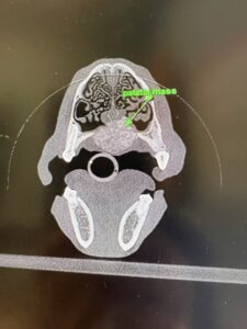
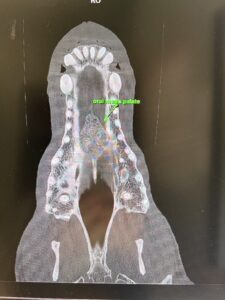
Figures 1 and 2: CT images pre-op
Initial surgery involved completely removing the hard palate including PM2.3.4 bilaterally. Bilateral advancement flap closure resulted in 75% healing at two weeks, but a fistula between the oral and nasal cavities persisted. In order to achieve complete closure, a lip rotational flap was used to reach across the palate and provide a thick, robust flap. The lip was then closed, front to back, and a tunnel created to keep the blood supply intact.
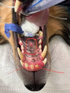
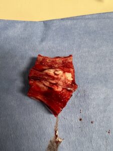
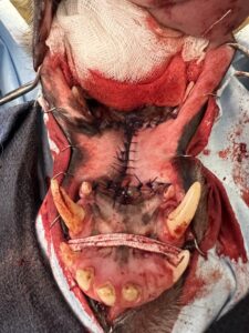
Figures 3, 4, and 5: Original surgery
At two weeks, this tunnel was severed and the result was complete closure of the defect with minimal cosmetic change to his appearance. His maxillary lips are slightly tucked in, and there is mild tension on the left side of his maxillary lip. The biopsy confirmed a MLO = multilobular osteosarcoma.
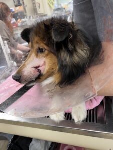
Figures 6 and 7: Two weeks post flap creation
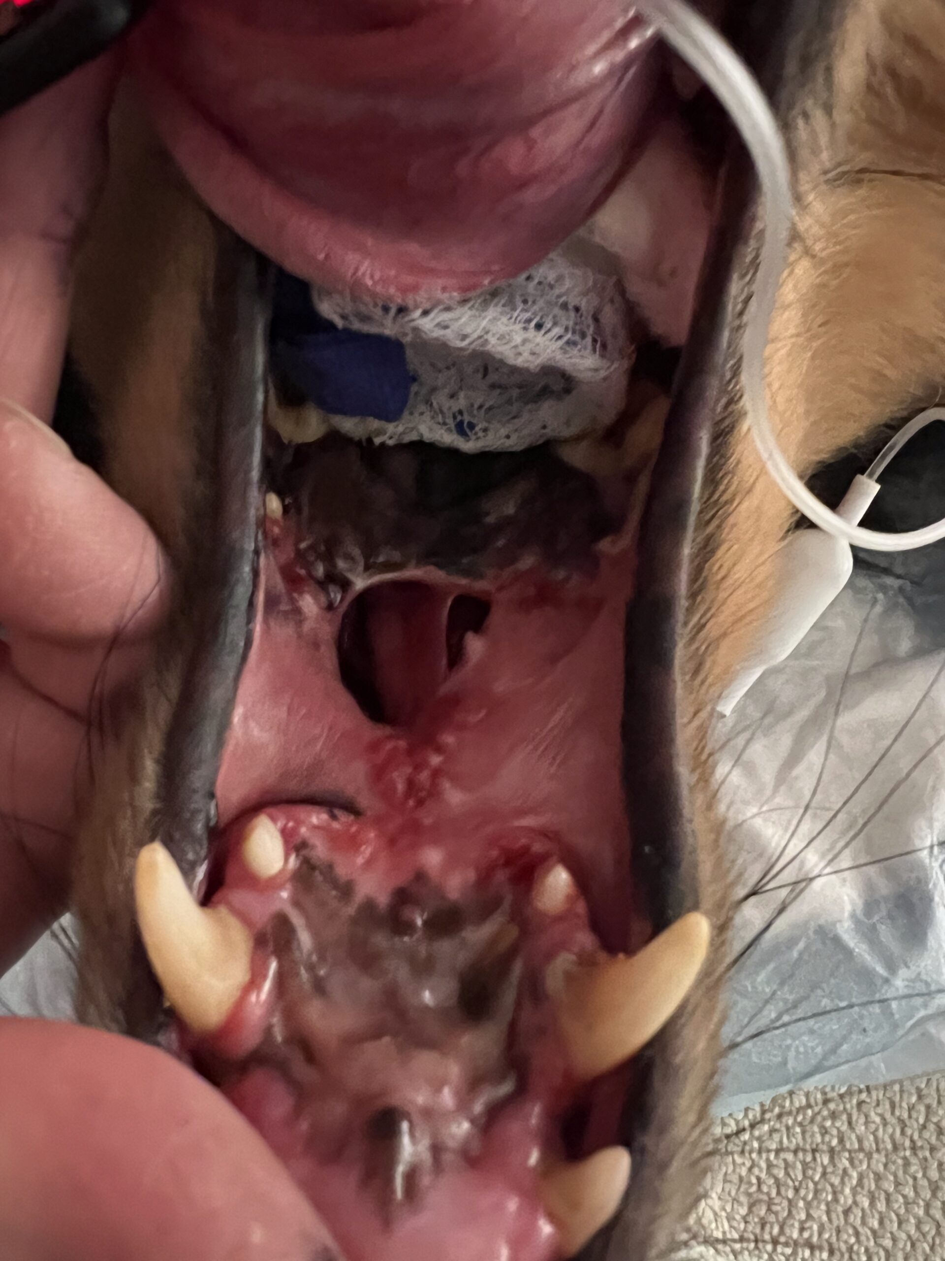
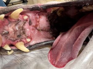
Figure 8: Partial dehiscence at first two week recheck
His prognosis is excellent with a long term remission or cure being reasonable expectations. No further treatment is recommended at this time and he will be monitored with annual CT scans for 3 years.
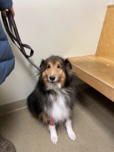
Figure 9: Healed appearance
Authored by: Marta Agrodnia, DVM, DACVS-SA (Surgery)